Examination of Arterial Pulse is a part of Cardiovascular System examination
Definition of Arterial Pulse
Arterial pulse is defined as the rhythmic expansion of the arterial wall due to transmission of pressure waves along the walls of the arteries, which are produced during each ventricular systole.
Importance of Examination of Arterial Pulse
It provides information regarding:
- Working condition of the heart
- Circulatory state and hemodynamics (blood volume, blood pressure)
- Condition of the blood vessels
- State of autonomic activity at that moment
- Mental state of the subject
- State of body metabolism and temperature
Why radial artery is chosen for Examination of Arterial Pulse?
The routine examination of arterial pulse is done on this
artery because:
- It is conveniently accessible as it is located in an exposed part of the body.
- The artery lies over the hard surface on the lower end of the radius.
Principle
With each ventricular contraction, not only is the blood
pumped into the aorta but also pressure waves generated are transmitted along
the walls of the vessels. These pressure waves expand the arterial wall and the
expansion is palpated as pulse.
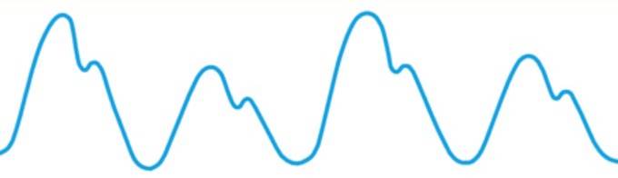
Arterial pulse wave Graph
Each pulse wave has a
- Percussion Wave/Anacrotic Limb
- Dicrotic Notch

Percussion Wave/Anacrotic Limb
- Sharp upstroke
- Due to expansion of the artery during ventricular systole: corresponds to the maximum ejection phase
Dicrotic Notch and Wave
- Seen on the descending limb.
- Negative wave, is due to recoil of the elastic aorta that causes the blood column to momentarily sweep back towards the heart. The reverse flow closes the aortic valve and rebounds from it to cause the positive dicrotic wave.
The radial artery is palpated with the tips of three fingers
compressing the vessel against the styloid process of radius.
The subject’s forearm should be slightly pronated and the
wrist slightly flexed.

Observation:
Parameters to check
- Rate (per min.)
- Rhythm
- Volume
- Character
- Condition of vessel wall
- Equality on both sides
- Radio-femoral delay
- Other peripheral pulses
1) Rate of Pulse
- Count the rate of the pulse (after calming the subject).
- Count the pulse completely for one minute.
- Take 3 readings and average them.
- Compare with heart beat for any pulse deficit.
- Pulse rate can never be more than the heart rate.
 |
Examination
of Arterial Pulse: Rate observation |
- Normal pulse rate 60-100/min.
- Sympathetic ↑, Parasympathethic ↓
- >100: Tachycardia, <60: Bradycardia
- Higher in children, low in old age
- Increases during deep inspiration and decreases during deep expiration.

2) Rhythm
- Spacing order at which successive pulse waves are felt
- Constant spacing: Regular, otherwise irregular
- Irregular pulse may have fixed pattern of irregularity (irregularly regular) or irregularity may not have any pattern (irregularly irregular)
- Regular/Irregularly regular/Irregularly irregular
 |
Examination
of Arterial Pulse: Rhythm observation |
- Due to generation of impulse from ectopic focus: ectopic beat
Respiratory sinus arrhthymia
- Irregularity of pulse rate constantly changes with respiration
- The pulse rate increases during deep inspiration and decreases during deep expiration: sinus arrhythmia: due to irradiation of impulses from the inspiratory center to the cardiac center.
Irregularly irregular pulse
- Atrial fibrillation
Irregularity associated with heart blocks: block in
conductivity
- Partial heart block with dropped beat
- Atrial flutter with irregular block
Pulse deficit
- Difference between heart rate and pulse rate
- Atrial fibrillation >10
- Heart blocks < 10
3) Volume
- Degree of expansion of the arterial walls
- Normal and equal on both sides
- Gives an indication of stroke volume of left ventricle
 |
Examination
of Arterial Pulse: Volume observation |
- Rough guide to pulse pressure
- Pulse pressure = SBP – DBP
- SBP depends on stroke volume
- DBP depends on compliance of the arteries
- Represents the force that the heart generates each time it contracts
- Low volume and High volume pulse
Low volume pulse:
- Pulsus parvus
- Aortic stenosis
- Obstructive cardiomyopathy
- Pericardial effusion
- Constrictive pericarditis
- Pulmonary stenosis
- Tight mitral stenosis
- Shock
High volume pulse:
- Pulsus magnus (Widening of pulse pressure)
- Aortic incompetence
- Thyrotoxicosis
- Patent ductus arteriosus
- Beriberi
- Anaemia
- Fever
- Old age
- Exercise

4) Character
Character of a pulse is said to be normal when no
abnormalities are detected either in rate, rhythm or volume of the pulse.
 |
Examination
of Arterial Pulse: Charecter observation |
Abnormal pulses
Anacrotic pulse
- Anadicrotic pulse
- Two upbeats
- Upstroke is slow and sloping
- Aortic stenosis

Dicrotic pulse
- Twice beating pulse
- Febrile states: typhoid fever

Water Hammer pulse
- Collapsing pulse or Corrigan’s pulse
- Aortic regurgitation
- Rapid upstroke and rapid downstroke
- Dicrotic notch is absent

Pulsus Bisferiens
- Combination of low rising pulse and collapsing pulse
- Aortic stenosis with aortic incompetence

- Constrictive pericarditis
- Pericardial effusion
Less common causes
- Emphysema
- Asthma
- Massive pleural effusion
- A mass in the thorax
- Advanced right ventricular failure
Pulsus Paradoxus
- Misnomer, nothing paradoxical
- Volume of pulse decrease during inspiration and increase during expiration

Pulsus alterans
- Regular alternating strong and weak beats
- Left ventricular failure, Toxic carditis
5) Condition of the arterial wall
- Young individuals: arterial wall is soft and elastic and not palpable
- Elderly: arterial wall is palpable, hard and may be tortuous: Due to thickening by atherosclerosis & arteriosclerosis

 |
Examination
of Arterial Pulse: Condition of the vessel wall observation |
6) Equality on both sides

7) Radio-radial or Radio-femoral delay
Radio-radial delay:
- Coarctation of the aorta proximal to origin of left subclavian artery
Radio-femoral delay:
- Coarctation of the aorta distal to origin of left subclavian artery

 |
Examination
of Arterial Pulse: Equality on both sides and radiofemoral delay
observation |
- Superficial temporal
- Carotid
- Brachial
- Femoral
- Popletial
- Posterior tibial
- Doralis pedis

 |
| Examination of Arterial Pulse: Other peripheral pulse observation |
Report:
On clinical examination of radial pulse of the subject _______ aged ___ yrs., the rate is ___ bpm., rhythm is regular, volume and character are normal. The vessel wall is not palpable because it is soft and elastic. There is no radio-radial as well as radio-femoral delay and equality is maintained on both the sides. Other peripheral pulses are felt and appear normal as well.